Gå til hovedinnhold
Havforskningsinstituttet
HI
English
<- Tilbake
Søk
Søk
Søk
View page in English
Figur
Fra rapporten:
Kunnskapsstøtte om miljøeffekter av kobber
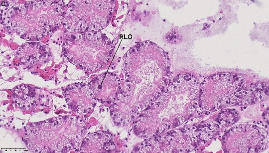
Figure 2. a) Debris in the gut, b) The same specimen as in figure a) examined under darkfield polarized interference microscope (Leica), C) RLO in the digestive tubule. NDP view 2, 40x (HAMATSU Photonics)